Anatomy Of Chest And Ribs : Xiphoid Process of Sternum / On the standard left lateral chest radiograph, the right ribs are projected behind the left and appear.
byAdmin-
0
Anatomy Of Chest And Ribs : Xiphoid Process of Sternum / On the standard left lateral chest radiograph, the right ribs are projected behind the left and appear.. The who manual of diagnostic imaging: This chapter is an abbreviated review of thoracic anatomy as seen on chest radiographs and computed tomography. Ribs eight to ten are the false ribs and are connected to the sternum indirectly via the cartilage of the rib clinical notes. As part of the bony thorax, the ribs protect the internal thoracic organs. Related posts of chest bone anatomy.
Radiographic anatomy and interpretation of chest and the pulmonary system. Normal anatomic structures are labeled on posteroanterior (pa) and lateral chest radiographs (figs. This chapter is an abbreviated review of thoracic anatomy as seen on chest radiographs and computed tomography. Insert contains images of a typical rib and the first rib. Continue scrolling to read more below.
Rib cage pain: 6 possible causes from www.medicalnewstoday.com This chapter is an abbreviated review of thoracic anatomy as seen on chest radiographs and computed tomography. Finally, it describes the muscles that cause the motion in the chest wall. Basic rib anatomy consists of a head, neck, tubercle. The bones of the chest and upper back combine to form the strong protective rib cage around the vital thoracic organs such as the heart and. How these parts interrelate through joints is described also. What are the features of ribs? Normal anatomic structures are labeled on posteroanterior (pa) and lateral chest radiographs (figs. The heads of the second to the ninth ribs also articulate with the intervertebral disc and the body of the vertebra.
Continue scrolling to read more below.
Spiral ct of thoracic inlet. Normal anatomic structures are labeled on posteroanterior (pa) and lateral chest radiographs (figs. Continue scrolling to read more below. It discusses the specific anatomy of the ribs and costal cartilages, along with the sternum. Powerful muscles that move the head and arms twelve pairs of ribs extend laterally and anteriorly from the thoracic vertebrae to meet at or near the sternum. Learn about chest anatomy with free interactive flashcards. ■ identify the basic anatomy seen on a chest radiograph. Ribs are divided into two basic groups the true ribs consist of 8 ribs, each on the left and right sides of the chest wall. This type of ct scan uses a lower radiation level than a conventional. How these parts interrelate through joints is described also. ■ describe the anatomical relationships of various organs in the chest. Identify the following structures on chest ct a good radiologist knows the anatomy because knowing where structures normally live and recognizing the location of an abnormality helps to make or narrow the differential diagnosis. The bones of the chest and upper back combine to form the strong protective rib cage around the vital thoracic organs such as the heart and.
Posteriorly, the heads of the ribs interdigitate with the vertebrae and are numbered according to the inferior vertebra. They also have a role in ventilation; Chest blunt trauma (cbt) and the resultant rib fractures often lead to thoracic collapse. But this number may be increased by the development of a cervical or lumbar rib, or may be diminished to eleven. The rib cage surrounds the lungs and the heart, serving as an important means of bony protection for these vital organs.
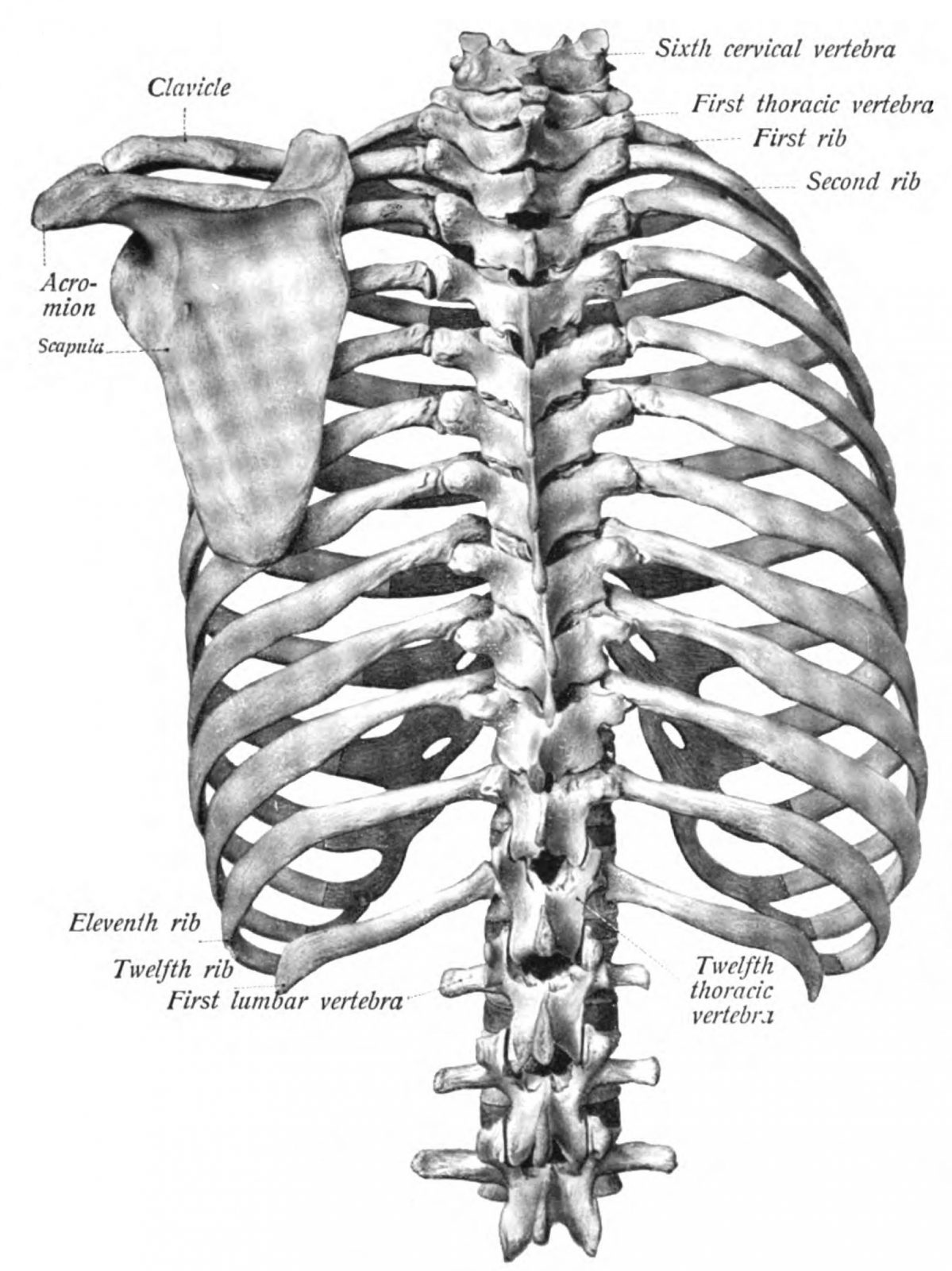
Anatomy of the Thorax → Thoracic Vertebral Column - Meddists from meddists.com As part of the bony thorax, the ribs protect the internal thoracic organs. Spiral ct of thoracic inlet. True, false and floating ribs are denoted. It discusses the specific anatomy of the ribs and costal cartilages, along with the sternum. The bones of the chest and upper back combine to form the strong protective rib cage around the vital thoracic organs such as the heart and. Insert contains images of a typical rib and the first rib. Anatomy is to physiology as geography is to history: ■ describe the anatomical relationships of various organs in the chest.
They are twelve in number on either side;
Related posts of chest bone anatomy. As part of the bony thorax, the ribs protect the internal thoracic organs. Ribs eight to ten are the false ribs and are connected to the sternum indirectly via the cartilage of the rib clinical notes. The pectoralis minor is a thin, triangular muscle that is found underneath the pectoralis major. Related online courses on physioplus. Learn about each muscle, their locations & functional anatomy. Learn about chest anatomy with free interactive flashcards. The spectrum of these rare anomalies includes unilateral absence, absence of cartilage, separation of cartilage and rib, combined skandalakis' surgical anatomy: The embryologic and anatomic basis of modern surgery. Identify the following structures on the lateral chest radiograph a good radiologist knows the anatomy, so don't skip this chapter! These true ribs are also numerically known as the 1st, 2nd, 3rd, 4th, 5th, 6th, 7th, and the 8th ribs. O bones—spine, ribs, clavicles, scapulae, humeri. How these parts interrelate through joints is described also.
Surface anatomy of anterior chest wall. ■ describe the anatomical relationships of various organs in the chest. The spectrum of these rare anomalies includes unilateral absence, absence of cartilage, separation of cartilage and rib, combined skandalakis' surgical anatomy: Respiratory muscle training online course: This chapter is an abbreviated review of thoracic anatomy as seen on chest radiographs and computed tomography.
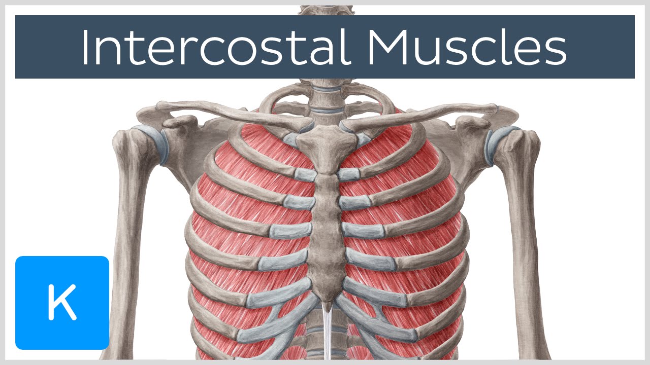
Intercostal Muscles - Function, Area & Course - Human ... from i.ytimg.com The heads of the second to the ninth ribs also articulate with the intervertebral disc and the body of the vertebra. Paschalides medical publications, 2004, with. Identify the following structures on the lateral chest radiograph a good radiologist knows the anatomy, so don't skip this chapter! The rib cage also anchors the bones of the head, neck, shoulders, and arms to the trunk of the body. Manubrium anteriorly, rib 1 laterally, thoracic vertebrae post… xiphoid process anteriorly, costal cartilages 7 to 10 and rib… Powerful muscles that move the head and arms twelve pairs of ribs extend laterally and anteriorly from the thoracic vertebrae to meet at or near the sternum. As with all parts of the body, the anatomy and physiology of the chest wall are intimately intertwined. It attaches at the 3rd, 4th and 5th rib, and it reaches to.
What are the features of ribs?
Related online courses on physioplus. Bone on hand and foot diagram quiz. The purpose of this study was to explore the effect of. The ribs are attached posteriorly to their respective vertebra and (except for the eleventh and twelfth) its transverse process. The pectoralis minor is a thin, triangular muscle that is found underneath the pectoralis major. Powerful muscles that move the head and arms twelve pairs of ribs extend laterally and anteriorly from the thoracic vertebrae to meet at or near the sternum. Radiographic anatomy and interpretation of chest and the pulmonary system. ■ identify the basic anatomy seen on a chest radiograph. The first seven are connected behind with the vertebral column. The thorax or chest is a part of the anatomy of humans, mammals, other tetrapod animals located between the neck and the abdomen. Insert contains images of a typical rib and the first rib. As part of the bony thorax, the ribs protect the internal thoracic organs. Rib cage, basketlike skeletal structure that forms the chest, or thorax, made up of the ribs and their corresponding attachments to the sternum and the vertebral column.
The spectrum of these rare anomalies includes unilateral absence, absence of cartilage, separation of cartilage and rib, combined skandalakis' surgical anatomy: anatomy of chest. Each rib wraps around the lung and descends approximately 3 to 5 inches.